


Bio-Imaging Facility
Confocal microscopy is an optical imaging technique for increasing optical resolution and contrast of a micrograph by means of using a spatial pinhole to block out-of-focus light in image formation. Capturing multiple two-dimensional images at different depths in a sample enables the reconstruction of three-dimensional structures within an object. At the NCCS - Bio-Imaging Facility, graduate and postdoctoral research fellows can be trained in microscope research techniques including advanced light microscopy, confocal microscopy, digital image processing of microscope images, and related laboratory techniques. Computer image processing and analysis is taught individually. In addition, the Facility offers workshops related to Microscopy and training programs are designed to train the students in modern and classical methods of preparing microscope slides from time to time. Staff members demonstrate the correct use of the instruments, train students in the microscopic techniques required for successful cell biological research and computer image processing and analysis.

- Dr. Ashwini N. Atre Technical Officer C
- Mrs. Trupti P. Kulkarni Technical Officer A
INFORMATION FOR EXTERNAL USERS:
- The Charges for External Users are Rs. 2000 per hour + 18 % GST for academic users and Rs. 3000 per hour + 18 % GST for non-academic users.
User will be charged according to the microscope usage time. User can bring DVD or pen drive for collecting data/ images. Outsiders will be allowed to use NCCS Microscopy facility maximum of two hours per week per microscope. To book the slots contact us @ bioimaging@nccs.res.in 020-25708265 / 8260 before 11 AM on previous Friday.
SERVICES PROVIDED:
- Expert consultation is provided through the Facility In-Charge and our technical specialists.
- Confocal microscopes are selected for complementary functions.
- Equipment use is accessible through dedicated technicians / operators.
- All services are provided on chargeable basis.
- Assistance with data analysis can be customized to the needs of individual investigators and research projects.
- Image manipulation and enhancement using Photoshop, ImageJ, Fiji, Matlab and Cell Profiler, IrfanView.
- Assistance in image and data analysis related to morphometrics, particle counting, particle tracking, numeric analysis, statistical analysis, etc.
- Data backup and archiving.
Requisition form
MICROSCOPES AVAILABLE AT NCCS BIO-IMAGING FACILITY
All the confocal systems are inverted microscopes and have a wide range of lasers.
Leica SP5 II Confocal Microscope
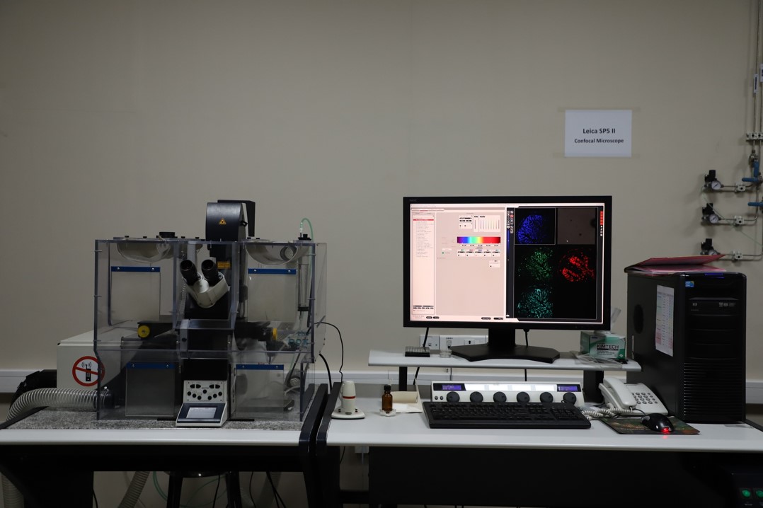
This is a high-end Broadband Confocal Laser Scanning Microscope with 3 PMT's, 2 Hybrid detectors and AOBS technology. The microscope is equipped with CO2 incubation chamber for live cell experiments as well as FRET, FRAP experiments and routine multi dye imaging. This is fully motorized, automated and computer controlled microscope Leica DMI 6000. Objectives available are 10x, 20x, 20x multi-immersion, 40x oil, 63x oil, 100x oil.
Olympus FV3000 Confocal Microscope
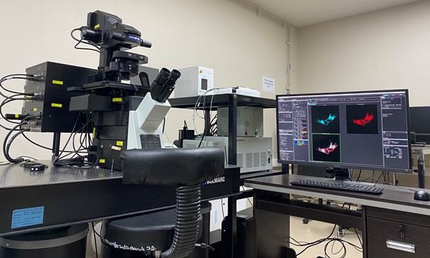
This is a high speed spectral confocal microscope for macro to microscopic imaging with 3D FRET, FRAP, Photo activation applications.It can attain high resolution nearly 140 nm or better (XY) and 350 nm in Z direction for at least two colours simultaneously with OSR (Olympus Super resolution- software based). Objectives are 1.25X to 60X. ZDC option (Z Drift Compensators) avoids minor vibrations during live imaging and retains the same focus position in real time for long term time lapse imaging. Images can be acquired at high speed or high scan format. It is equipped with a fully motorized XYstage inverted microscope with anti-vibration platform. The system has 4 detectors- 2 PMT detectors and 2 High sensitivity detectors (GaAsP) with possibility of 8 to 16 channels and dedicated transmitted light detector for DIC imaging.
Thermo CellInsight CX7 LZR - Confocal based High Content Screening System

This is a fast, laser-based, automated cellular imaging and analysis platform for quantitative microscopy and phenotypic screening. It is a 7-channel, laser-based illumination system with Software and laser-based autofocus for consistent scan times. The objectives range from 2x, 10x, 20x, 40x to 60x. Major applications are fluorescent, confocal and 5-channel brightfield imaging. It also has an onstage incubator for live cell imaging. It is highly compatible with standard microplates (6, 24, 48, 96, 384, 1536 wells). It is loaded with HCS Studio software for integrated data collection and analysis. The system provides powerful statistical method to carryout various bioapplications like- Autophagy assay, Cell viability assay, Kinetic assays, Apoptosis, DNA damage, Transfection efficiency, Cell toxicity and colony formation assays etc.
Zeiss LSM880 Confocal Microscope with AiryScan and ELYRA P.1

This imaging system comprises of AxioObserver 7 microscope with AiryScan and Elyra P.1 technology on the LSM 880. Airyscan is a 32-channel GaAsP-PMT area detector that collects a pinhole-plane image at every scan position. Each detector element functions as a single, very small pinhole. ELYRA P.1 localizes small structures and even single molecules achieving resolutions of 20nm laterally and 50nm axially.
Olympus SPIN SR 10 - Spinning Disk High Resolution Microscope

This is a Spinning disc confocal microscope equipped with YOKOGAWA W1- SoRa scan-unit. It can attain high resolution minimum 250 nm (XY) and 800 nm in Z. Super-resolution mode gives XY resolution of 120nm and Z of 540nm approximately. Objectives are 10X to 100X. ZDC option (Z Drift Compensators) avoids minor vibrations during live imaging and retains the same focus position in real time for long term time lapse imaging. Images can be acquired at high speed or high scan format. It is equipped with a fully motorized XY stage inverted microscope with anti-vibration platform. The system has 2 high-speed cameras for simultaneous imaging of atleast two dyes.
Contact Information

020-25708265/8260

bioimaging@nccs.res.in

Bio-Imaging Facility,
National Centre for Cell Science,
NCCS Complex, P.B. No. 40, Ganeshkhind Post,
S. P. Pune University Campus,
PUNE – 411 007,
Maharashtra State, India.
Fax: +91-20-2569 2259
Directions to Bio-Imaging Facility: Situated in NCCS New Building Ground Floor (Next to Reception)